Miquel Stoudmire
  • Home
  • Blog

Fallen Arches Causes, Indicators And Treatment Method

7/5/2017

 
Overview

Acquired Flat Foot

The condition known as ?fallen arches? is also referred to as flat feet or pes planus. It arises when the arch of the foot collapses to the point that the entire sole is flat to the ground. It is commonly seen in young children who have yet to develop the muscles in the sole of their feet. In most cases as a child develops and learns how to walk the finer intrinsic muscles that support the sole of the foot will develop normally and an arch forms. However in a small number of cases these muscles don?t form properly and thus neither does the arch. Fallen arches which are acquired in adulthood may be due to a number of factors.

Causes

There is a lack of normal arch development, probably due to inherent ligamentous laxity. Around 20% of adults have Pes planus. The majority have a flexible flat foot and no symptoms. However, if there is also heel cord contracture, there may be symptoms (see 'Contributing factors', below). Loss of support for the arch. Dysfunction of the tibialis posterior tendon, a common and important cause. Tear of the spring ligament (rare). Tibialis anterior rupture (rare). A neuropathic foot, e.g from diabetes, polio, or other neuropathies. Degenerative changes in foot and ankle joints. Inflammatory arthropathy, eg rheumatoid arthritis. Osteoarthritis. Fractures. Bony abnormalities, eg tarsal coalition.

Symptoms

Having flat feet can be painless and is actually normal in some people. But others with flat feet experience pain in the heel or arch area, difficulty standing on tiptoe, or have swelling along the inside of the ankle. They may also experience pain after standing for long periods of time or playing sports. Some back problems can also be attributed to flat feet.

Diagnosis

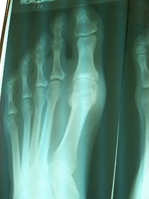
Your doctor will ask about your symptoms and medical history. A physical and foot exam will be done. Flat feet can be diagnosed by appearance. To determine if the foot is rigid, you may be asked to do some simple tasks.

no-foot-pain.com

Non Surgical Treatment

Normally, flat feet disappear by age six as the feet become less flexible and the arches develop. Only about 1 or 2 out of every 10 children will continue to have flat feet into adulthood. For children who do not develop an arch, treatment is not recommended unless the foot is stiff or painful. Shoe inserts won?t help your child develop an arch, and may cause more problems than the flat feet themselves. However, certain forms of flat feet may need to be treated differently. For instance, a child may have tightness of the heel cord (Achilles tendon) that limits the motion of his foot. This tightness can result in a flat foot, but it usually can be treated with special stretching exercises to lengthen the heel cord. Rarely, a child will have truly rigid flat feet, a condition that can cause problems.

Surgical Treatment

Adult Acquired Flat Foot

A better approach is to strengthen the weakened ligaments with Prolotherapy, supplemented by an arch support if the condition has existed for several years. Chronic pain is most commonly due to tendon and ligament weakness, or cartilage deterioration. The safest and most effective natural medicine treatment for repairing tendon, ligament and cartilage damage is Prolotherapy. In simple terms, Prolotherapy stimulates the body to repair painful areas. It does so by inducing a mild inflammatory reaction in the weakened ligaments and cartilage. Since the body heals by inflammation, Prolotherapy stimulates healing. Prolotherapy offers the most curative results in treating chronic pain. It effectively eliminates pain because it attacks the source: the fibro-osseous junction, an area rich in sensory nerves. What?s more, the tissue strengthening and pain relief stimulated by Prolotherapy is permanent.

Prevention

Strap the arches into the anatomically correct positions with athletic tape and leave them like this for some time. If the fallen arches are an issue with the muscular structure, this may give the muscles an opportunity to strengthen. This is definitely not a fallen arches cure all the time but it can help prevent it more times than not. Ask a doctor or physical therapists to show you how to do this taping. Find shoes that fit. This may require that you get your foot measured and molded to ensure that the shoe will fit. Shoes that are too big, too tight or too short, may not directly cause the fallen arches, but they can assist with the damage to the area. These shoes should have thick cushioning inside and have plenty of room for your toes. Walk without shoes as much as possible. Shoes directly assist with weakening and distorting the arches of the feet so going without shoes can actually help strengthen your arches and prevent fallen arches. Walking on hard and bumpy surfaces barefooted makes the muscles in your feet strengthen in order to prevent injury. It is a coping mechanism by your body. Insert heel cups or insoles into the shoes that you wear the most. Many people wear uncomfortable shoes to work and these are the same shoes that cause their arches the most problems. Inserting the heel cups and insoles into these shoes can prevent fallen arches from occurring. Many people place these inserts into all their shoes to ensure support. Ask a medical professional, either your doctor or a physical therapist, about daily foot exercises that may keep the arches stronger than normal. Many times, you can find exercises and stretches on the Internet on various websites. Curling your toes tightly and rotating your feet will help strengthen your longitudinal arches. Relax your feet and shake them for a minute or so before you do any arch exercises. This will loosen the muscles in your feet that stay tight due to normal daily activities. Wear rigid soled sandals whenever possible to provide a strong support for your arches. Wooden soled sandals are the best ones if available. Walk or jog on concrete as much as you can. This will create a sturdy support for your arches. Running or walking in sandy areas or even on a treadmill, does not give rigid support. Instead, these surfaces absorb the step, offering no support for arches.

After Care

Patients may go home the day of surgery or they may require an overnight hospital stay. The leg will be placed in a splint or cast and should be kept elevated for the first two weeks. At that point, sutures are removed. A new cast or a removable boot is then placed. It is important that patients do not put any weight on the corrected foot for six to eight weeks following the operation. Patients may begin bearing weight at eight weeks and usually progress to full weightbearing by 10 to 12 weeks. For some patients, weightbearing requires additional time. After 12 weeks, patients commonly can transition to wearing a shoe. Inserts and ankle braces are often used. Physical therapy may be recommended. There are complications that relate to surgery in general. These include the risks associated with anesthesia, infection, damage to nerves and blood vessels, and bleeding or blood clots. Complications following flatfoot surgery may include wound breakdown or nonunion (incomplete healing of the bones). These complications often can be prevented with proper wound care and rehabilitation. Occasionally, patients may notice some discomfort due to prominent hardware. Removal of hardware can be done at a later time if this is an issue. The overall complication rates for flatfoot surgery are low.

Heel Pain All You Ought To Understand Heel Painfulness

7/1/2017

 
Overview

Heel Pain

Heel pain is often a symptom caused by one of two conditions: Plantar Fasciitis or Achilles Tendonitis. Most commonly, heel pain experienced at the bottom of the heel is caused by plantar fasciitis. Heel pain may become so severe for some that just putting weight on their feet first thing in the morning is excruciating. Walking or running may feel completely out of the question.

Causes

Heel pain is a common symptom that has many possible causes. Although heel pain sometimes is caused by a systemic (body-wide) illness, such as rheumatoid arthritis or gout, it usually is a local condition that affects only the foot. The most common local causes of heel pain include Plantar fasciitis. lantar fasciitis is a painful inflammation of the plantar fascia, a fibrous band of tissue on the sole of the foot that helps to support the arch. Plantar fasciitis occurs when the plantar fascia is overloaded or overstretched. This causes small tears in the fibers of the fascia, especially where the fascia meets the heel bone. Plantar fasciitis may develop in just about anyone but it is particularly common in the following groups of people: people with diabetes, obese people, pregnant women, runners, volleyball players, tennis players and people who participate in step aerobics or stair climbing. You also can trigger plantar fasciitis by pushing a large appliance or piece of furniture or by wearing worn out or poorly constructed shoes. In athletes, plantar fasciitis may follow a period of intense training, especially in runners who push themselves to run longer distances. People with flat feet have a higher risk of developing plantar fasciitis. Heel spur. heel spur is an abnormal growth of bone at the area where the plantar fascia attaches to the heel bone. It is caused by long-term strain on the plantar fascia and muscles of the foot, especially in obese people, runners or joggers. As in plantar fasciitis, shoes that are worn out, poorly fitting or poorly constructed can aggravate the problem. Heel spurs may not be the cause of heel pain even when seen on an X-ray. In fact, they may develop as a reaction to plantar fasciitis. Calcaneal apophysitis, n this condition, the center of the heel bone becomes irritated as a result of a new shoe or increased athletic activity. This pain occurs in the back of the heel, not the bottom. Calcaneal apophysitis is a fairly common cause of heel pain in active, growing children between the ages of 8 and 14. Although almost any boy or girl can be affected, children who participate in sports that require a lot of jumping have the highest risk of developing this condition. Bursitis. ursitis means inflammation of a bursa, a sac that lines many joints and allows tendons and muscles to move easily when the joint is moving. In the heel, bursitis may cause pain at the underside or back of the heel. In some cases, heel bursitis is related to structural problems of the foot that cause an abnormal gait (way of walking). In other cases, wearing shoes with poorly cushioned heels can trigger bursitis. Pump bump. his condition, medically known as posterior calcaneal exostosis, is an abnormal bony growth at the back of the heel. It is especially common in young women, in whom it is often related to long-term bursitis caused by pressure from pump shoes. Local bruises. ike other parts of the foot, the heel can be bumped and bruised accidentally. Typically, this happens as a "stone bruise," an impact injury caused by stepping on a sharp object while walking barefoot. Achilles tendonitis. n most cases, Achilles tendonitis (inflammation of the Achilles tendon) is triggered by overuse, especially by excessive jumping during sports. However, it also can be related to poorly fitting shoes if the upper back portion of a shoe digs into the Achilles tendon at the back of the heel. Less often, it is caused by an inflammatory illness, such as ankylosing spondylitis (also called axial spondylarthritis), reactive arthritis, gout or rheumatoid arthritis. Trapped nerve. ompression of a small nerve (a branch of the lateral plantar nerve) can cause pain, numbness or tingling in the heel area. In many cases, this nerve compression is related to a sprain, fracture or varicose (swollen) vein near the heel.

Symptoms

See your doctor immediately if you have Severe pain and swelling near your heel. Inability to bend your foot downward, rise on your toes or walk normally. Heel pain with fever, numbness or tingling in your heel. Severe heel pain immediately after an injury. Schedule an office visit if you have. Heel pain that continues when you're not walking or standing. Heel pain that lasts more than a few weeks, even after you've tried rest, ice and other home treatments.

Diagnosis

To arrive at a diagnosis, the foot and ankle surgeon will obtain your medical history and examine your foot. Throughout this process the surgeon rules out all the possible causes for your heel pain other than plantar fasciitis. In addition, diagnostic imaging studies such as x-rays or other imaging modalities may be used to distinguish the different types of heel pain. Sometimes heel spurs are found in patients with plantar fasciitis, but these are rarely a source of pain. When they are present, the condition may be diagnosed as plantar fasciitis/heel spur syndrome.

Non Surgical Treatment

Most heel pain is caused by a combination of poor biomechanics, or muscle weakness or tightness. The good news is that heel pain can be effectively managed once the cause is identified. Most heel pain can be successfully treated via pain and pressure relief techniques, biomechanical correction eg orthotics, taping, foot posture exercises, muscle stretches and massage, lower limb muscle strengthening, proprioceptive and balance exercises to stimulate your foot intrinsic muscles. If you feel that your footwear or sports training schedule are potentially causing your heel pain, then we recommend that you seek the advice of a sports physiotherapist, podiatrist or trained footwear specialist (not just a shop assistant) to see if your shoe is a match for your foot; or discuss your training regime to see if you are doing too much. Heel pain and injury are extremely common. With accurate assessment and early treatment most heel pain injuries respond extremely quickly to physiotherapy allowing you to quickly resume pain-free and normal activities of daily living. Please ask you physiotherapist for their professional treatment advice.

Surgical Treatment

Although most patients with plantar fasciitis respond to non-surgical treatment, a small percentage of patients may require surgery. If, after several months of non-surgical treatment, you continue to have heel pain, surgery will be considered. Your foot and ankle surgeon will discuss the surgical options with you and determine which approach would be most beneficial for you. No matter what kind of treatment you undergo for plantar fasciitis, the underlying causes that led to this condition may remain. Therefore, you will need to continue with preventive measures. Wearing supportive shoes, stretching, and using custom orthotic devices are the mainstay of long-term treatment for plantar fasciitis.

heel cups for heel pain

Prevention

Heel Pain

Heel pain is commonly caused from shoes that do not fit properly. In addition, shoes need to have ample cushioning and support, particularly through the heel, ball of the foot, and arch. Shoes should also be replaced if they become too worn. One sure sign of wear and tear is overly worn areas of a shoe's insoles. If the heel or ball of the foot is particularly worn, damage could easily occur since the bottom of the foot is not getting the cushioning it needs.

Limb-Length Discrepancy After Hip Arthroplasty

6/29/2017

 
Overview

Leg length discrepancy is the difference in lengths of an individual?s legs. This difference may be anatomical or may be due to scoliosis, trauma/injury, arthritis, overpronation (collapse) of one foot, bowing of one leg or unequal bowing, surgery (hip or knee replacement), pelvic tilting or ageing. The difference can also be functional caused by differing forces of the soft tissues, such as weakness in muscle tissue on one side, or a weakness/tightness in joint tissue. A difference in leg lengths also results when running on indoor banked tracks, beaches and banked streets and side walks (for drainage). Many people have a measurable difference in their leg lengths which is compensated for by their bodies. As we age this compensation does not work as well. An x-ray and physical measurements will define the discrepancy and the adjustment needed.Leg Length Discrepancy

Causes

A patient?s legs may be different lengths for a number of reasons, including a broken leg bone may heal in a shorter position, particularly if the injury was severe. In children, broken bones may grow faster for a few years after they heal, resulting in one longer leg. If the break was near the growth center, slower growth may ensue. Children, especially infants, who have a bone infection during a growth spurt may have a greater discrepancy. Inflammation of joints, such as juvenile arthritis during growth, may cause unequal leg length. Compensation for spinal or pelvic scoliosis. Bone diseases such as Ollier disease, neurofibromatosis, or multiple hereditary exostoses. Congenital differences.

Symptoms

Patients with significant lower limb length discrepancies may walk with a limp, have the appearance of a curved spine (non-structural scoliosis), and experience back pain or fatigue. In addition, clothes may not fit right.

Diagnosis

The doctor carefully examines the child. He or she checks to be sure the legs are actually different lengths. This is because problems with the hip (such as a loose joint) or back (scoliosis) can make the child appear to have one shorter leg, even though the legs are the same length. An X-ray of the child?s legs is taken. During the X-ray, a long ruler is put in the image so an accurate measurement of each leg bone can be taken. If an underlying cause of the discrepancy is suspected, tests are done to rule it out.

Non Surgical Treatment

A personalized approach to leg length discrepancy treatment works best for each patient. Your child's doctor will consider many factors when deciding on a course of treatment for this condition. Such factors can include your child's age, extent of the leg length discrepancy, medical history, how your child tolerates certain treatments and procedures, your child's health and prognosis, growth rate, and parental preferences. There is no cookie cutter treatment for each child, or even each centimeter of leg difference. Once all of these factors have been carefully considered, your child's physician will come up with an appropriate course of care. In situations of a very small leg length discrepancy, no treatment, only periodic medical evaluation, may be necessary.

Leg Length

what is a heel raise?

Surgical Treatment

Your child will be given general anesthetic. We cut the bone and insert metal pins above and below the cut. A metal frame is attached to the pins to support the leg. Over weeks and months, the metal device is adjusted to gradually pull the bone apart to create space between the ends of the bones. New bone forms to fill in the space, extending the length of the bone. Once the lengthening process is completed and the bones have healed, your child will require one more short operation to remove the lengthening device. We will see your child regularly to monitor the leg and adjust the metal lengthening device. We may also refer your child to a physical therapist to ensure that he or she stays mobile and has full range of motion in the leg. Typically, it takes a month of healing for every centimeter that the leg is lengthened.

Coping with Mortons Neuroma

6/5/2017

 
Overview

Morton neuromaA Morton?s Neuroma is actually incorrectly termed, with the name suggesting it is a tumour or growth. Rather than a true neuroma it is actually what is called a perineural fibrosis, which means that over time the sheath surrounding the nerve becomes irritated, inflamed, and forms a thickened scar tissue.

Causes

A Morton?s Neuroma are a result of complex biomechanical changes that occur in your feet. There are a number of theories as to the exact cause of the scarring and thickening, but it basically boils down to overload of the tissue structure. The body lays down scar tissue to try to protect the overloaded structure. Tight-fitting shoes may exacerbate a Morton?s Neuroma. Shoes such as high heels and shoes with tight toe boxes (eg womens fashion shoes and cowboy boots) are particularly damaging to the toes. These shoes have a sloping foot bed and a narrow toe box. The slope causes the front of the foot to bear your weight. The angle of the toe box then squeezes your toes together. Footwear is not the only cause of a Morton?s Neuroma. Injuries to the foot can also be a factor in developing the condition by changing your foot biomechanics. Poor foot arch control leading to flat feet or foot overpronation does make you biomechanically susceptible to a neuroma.

Symptoms

Patients will feel pain that worsens with walking, particularly when walking in shoes with thin soles or high heels. Also, anything that squeezes the metatarsal heads together may aggravate symptoms, such as narrow shoes. A patient may feel the need to remove the shoe and rub the foot to soothe the pain.

Diagnosis

You might first seek advice from your family doctor about your foot pain. He or she may refer you to a doctor or surgeon who specializes in foot disorders. Before your appointment, you may want to write a list of answers to the following questions. When did your symptoms begin? Did your symptoms begin gradually or suddenly? What type of footwear do you wear for work? Do you participate in sports? If so, what types in particular? What medications and supplements do you take regularly? Your doctor may ask some of the following questions. Is the pain worse in certain pairs of shoes? Does any type of activity ease the pain or worsen it? Are you having pain in any other part of your body?

Non Surgical Treatment

In developing a treatment plan, your foot and ankle surgeon will first determine how long you?ve had the neuroma and evaluate its stage of development. Treatment approaches vary according to the severity of the problem. For mild to moderate neuromas, treatment options may include Padding techniques provide support for the metatarsal arch, thereby lessening the pressure on the nerve and decreasing the compression when walking. Placing an icepack on the affected area helps reduce swelling. Custom orthotic devices provided by your foot and ankle surgeon provide the support needed to reduce pressure and compression on the nerve. Activities that put repetitive pressure on the neuroma should be avoided until the condition improves. Wear shoes with a wide toe box and avoid narrow-toed shoes or shoes with high heels. Oral nonsteroidal anti-inflammatory drugs (NSAIDs), such as ibuprofen, may be recommended to reduce pain and inflammation. Treatment may include injections of cortisone, local anesthetics or other agents.Morton

Surgical Treatment

Interdigital neurectomy (removal of the diseased nerve) in right hands, should give satisfactory results almost all the time. Some of the reasons behind failure is when not enough nerve is dissected, mistakes in initial diagnosis, or bad handling of adjacent nerves, tendons and joint capsules during the operation. It is very common and acceptable to have some numbness in the area where the nerve used to be. This never causes any discomfort and often gets better in few years. It is crucial to address the biomechanical pathologies underlying the impingement of the nerve during and after the surgery.

Prevention

The best way to prevent a neuroma is by avoiding the things that cause them. Review your risk factors. If relatives have had similar problems, or if you know that you pronate or have any problem with the mechanics of your foot, talk with a podiatric physician about the correct types of shoes and/or orthoses for you. If you are not sure whether you have such a problem, the podiatric professional can analyze your foot, your stride and the wear pattern of your shoes, and give you an honest evaluation. Remember, though, that sometimes neuromas, like other conditions, can develop for no discernible reason. With this in mind, be good to your feet, and be aware of any changes or problems. Don?t wait to report them.

Leg Length Discrepancy And Shoe Lifts

3/3/2016

 
There are actually two different types of leg length discrepancies, congenital and acquired. Congenital implies you are born with it. One leg is structurally shorter in comparison to the other. As a result of developmental phases of aging, the brain senses the stride pattern and identifies some variation. The human body usually adapts by tilting one shoulder over to the "short" side. A difference of less than a quarter inch isn't blatantly abnormal, doesn't need Shoe Lifts to compensate and usually won't have a serious effect over a lifetime.

Leg Length Discrepancy Shoe Lift

Leg length inequality goes typically undiagnosed on a daily basis, yet this issue is simply corrected, and can reduce quite a few instances of upper back pain.

Treatment for leg length inequality typically involves Shoe Lifts. These are generally cost-effective, typically costing under twenty dollars, compared to a custom orthotic of $200 if not more. Differences over a quarter inch can take their toll on the spine and should probably be compensated for with a heel lift. In some cases, the shortage can be so extreme that it requires a full lift to both the heel and sole of the shoe.

Lumbar pain is the most prevalent health problem impacting people today. Over 80 million people suffer from back pain at some point in their life. It is a problem that costs businesses huge amounts of money each year as a result of time lost and output. Innovative and better treatment solutions are always sought after in the hope of lowering economic influence this issue causes.

Leg Length Discrepancy  <a href="http://tightidol5014.soup.io/tag/shoe%20lifts">Shoe Lifts</a>

People from all corners of the world suffer from foot ache as a result of leg length discrepancy. In these types of situations Shoe Lifts are usually of beneficial. The lifts are capable of relieving any discomfort and pain in the feet. Shoe Lifts are recommended by many qualified orthopaedic practitioners".

To be able to support the human body in a nicely balanced manner, your feet have got a very important job to play. Despite that, it is often the most neglected area of the human body. Some people have flat-feet meaning there may be unequal force exerted on the feet. This will cause other body parts like knees, ankles and backs to be impacted too. Shoe Lifts make sure that ideal posture and balance are restored.

Shoe Lifts For Leg Length Discrepancy

2/28/2016

 
There are actually not one but two different types of leg length discrepancies, congenital and acquired. Congenital implies that you are born with it. One leg is structurally shorter compared to the other. As a result of developmental stages of aging, the human brain senses the stride pattern and recognizes some variation. The human body typically adapts by tilting one shoulder to the "short" side. A difference of under a quarter inch is not really irregular, require Shoe Lifts to compensate and mostly doesn't have a serious effect over a lifetime.

Shoe Lift

Leg length inequality goes mainly undiscovered on a daily basis, yet this issue is very easily fixed, and can reduce many cases of back ache.

Therapy for leg length inequality typically involves Shoe Lifts. Many are low-priced, in most cases costing less than twenty dollars, compared to a custom orthotic of $200 and up. When the amount of leg length inequality begins to exceed half an inch, a whole sole lift is generally the better choice than a heel lift. This prevents the foot from being unnecessarily stressed in an abnormal position.

Low back pain is easily the most common ailment impacting people today. Over 80 million men and women are afflicted by back pain at some stage in their life. It's a problem that costs employers millions of dollars yearly due to time lost and output. New and more effective treatment solutions are always sought after in the hope of lowering economical impact this condition causes.

Shoe Lift

Men and women from all corners of the world suffer the pain of foot ache due to leg length discrepancy. In a lot of these situations Shoe Lifts can be of immense help. The lifts are capable of reducing any pain in the feet. Shoe Lifts are recommended by many certified orthopaedic practitioners".

So that they can support the human body in a balanced fashion, the feet have a crucial role to play. Irrespective of that, it is often the most neglected zone in the body. Some people have flat-feet meaning there is unequal force exerted on the feet. This will cause other areas of the body such as knees, ankles and backs to be affected too. Shoe Lifts ensure that suitable posture and balance are restored.

Are Shoe Lifts The Answer To Leg Length Discrepancy

2/26/2016

 
There are actually not one but two unique variations of leg length discrepancies, congenital and acquired. Congenital implies you are born with it. One leg is anatomically shorter compared to the other. Through developmental periods of aging, the human brain senses the walking pattern and identifies some difference. Our bodies usually adapts by tilting one shoulder over to the "short" side. A difference of under a quarter inch is not really irregular, does not need Shoe Lifts to compensate and in most cases won't have a profound effect over a lifetime.

Leg Length Discrepancy Shoe Lifts

Leg length inequality goes largely undiscovered on a daily basis, however this problem is very easily fixed, and can eradicate many cases of low back pain.

Treatment for leg length inequality commonly consists of Shoe Lifts. Most are very inexpensive, frequently costing under twenty dollars, compared to a custom orthotic of $200 or maybe more. When the amount of leg length inequality begins to exceed half an inch, a whole sole lift is generally the better choice than a heel lift. This prevents the foot from being unnecessarily stressed in an abnormal position.

Back pain is the most prevalent condition afflicting people today. Over 80 million men and women are afflicted by back pain at some stage in their life. It is a problem which costs employers vast amounts of money annually on account of time lost and output. New and more effective treatment methods are continually sought after in the hope of minimizing the economical influence this condition causes.

Leg Length Discrepancy Shoe Lift

People from all corners of the earth suffer the pain of foot ache due to leg length discrepancy. In most of these situations Shoe Lifts are usually of very helpful. The lifts are capable of easing any discomfort and pain in the feet. Shoe Lifts are recommended by numerous skilled orthopaedic doctors.

To be able to support the body in a well balanced fashion, the feet have a very important role to play. In spite of that, it's often the most overlooked area of the human body. Some people have flat-feet which means there is unequal force exerted on the feet. This will cause other body parts like knees, ankles and backs to be affected too. Shoe Lifts ensure that proper posture and balance are restored.

What Would Cause Severs Disease?

5/20/2015

 
Overview

Sever's Disease, also known as calcaneal apophysitis, is a disease of the growth plate of the heel bone (calcaneus) and is characterized by pain in the heel of a child's foot, typically brought on by some form of injury or minor trauma from sports participation. This condition is most common in children ages 9 to 14 and is frequently seen in active soccer, football, or baseball players. Sport shoes with cleats are also known to aggravate the condition. The disease can mimic Achilles tendonitis, an inflammation of the tendon attached to the back of the heel. A tight Achilles tendon contributes to Sever's Disease by pulling excessively on the growth plate of the heel bone. Feet that flatten out to excess (hyperpronate) are also prone to this problem, as this can involve extra torque on the calcaneus by the Achilles tendon.

Causes

Severs disease is often associated with a rapid growth spurt. As the bones get longer, the muscles and tendons become tighter as they cannot keep up with the bone growth. The point at which the achilles tendon attaches to the heel becomes inflamed and the bone starts to crumble (a lot like osgood schlatters disease of the knee). Tight calf muscles may contribute as the range of motion at the ankle is reduced resulting in more strain on the achilles tendon. Sever's disease is the second most common injury of this type which is known as an apophysitis.

Symptoms

The typical clinical presentation is an active child (aged 9-10 years) who complains of pain at the posterior heel that is made worse by sports, especially those involving running or jumping. The onset is usually gradual. Often, the pain has been relieved somewhat with rest and consequently has been patiently monitored by the patient, parents, coaches, trainers, and family physicians, in the expectation that it will resolve. When the pain continues to interfere with sports performance and then with daily activities, further consultation is sought. It should be kept in mind that failure to instruct patients and parents that continual pain, significant swelling or redness, and fever are not signs of Sever disease and therefore require further evaluation could result in failure to diagnose a condition with much more serious long-term consequences.

Diagnosis

Sever?s disease can be diagnosed based on the symptoms your child has. Your child?s doctor will conduct a physical examination by squeezing different parts of your child?s foot to see if they cause any pain. An X-ray may be used to rule out other problems, such as a broken bone or fracture.

Non Surgical Treatment

Decreasing or stopping sport is necessary until the pain reduces. Let pain be your guide, as it decreases you can slowly return to all activities. To help settle inflammation use an ice pack or rub an ice cube over the

painful area for 5 minutes daily whilst pain persists. Wearing supportive trainers during the day can help to soften the impact of walking on the heel. Encourage a normal pattern of walking. Complete the stretches below every day and before and after activity until your symptoms settle.

Surgical Treatment

The surgeon may select one or more of the following options to treat calcaneal apophysitis. Reduce activity. The child needs to reduce or stop any activity that causes pain. Support the heel. Temporary shoe inserts or custom orthotic devices may provide support for the heel. Medications. Nonsteroidal anti-inflammatory drugs (NSAIDs), such as ibuprofen, help reduce the pain and inflammation. Physical therapy. Stretching or physical therapy modalities are sometimes used to promote healing of the inflamed issue. Immobilization. In some severe cases of pediatric heel pain, a cast may be used to promote healing while keeping the foot and ankle totally immobile. Often heel pain in children returns after it has been treated because the heel bone is still growing. Recurrence of heel pain may be a sign of calcaneal apophysitis, or it may indicate a different problem. If your child has a repeat bout of heel pain, be sure to make an appointment with your foot and ankle surgeon.

Which Are The Principal Causes Of Adult Aquired FlatFeet ?

3/28/2015

 
Overview

Adult acquired flatfoot is one of the most common problems affecting the foot and ankle. Treatment ranges from nonsurgical methods, such as orthotics and braces to surgery. Your doctor will create a treatment plan for you based on what is causing your AAFD.Acquired Flat Feet


Causes

Posterior tibial tendon dysfunction is the most common cause of acquired adult flatfoot deformity. There is often no specific event that starts the problem, such as a sudden tendon injury. More commonly, the tendon becomes injured from cumulative wear and tear. Posterior tibial tendon dysfunction occurs more commonly in patients who already have a flat foot for other reasons. As the arch flattens, more stress is placed on the posterior tibial tendon and also on the ligaments on the inside of the foot and ankle. The result is a progressive disorder.


Symptoms

Some symptoms of adult acquired flat foot are pain along the inside of the foot and ankle, pain that increases with activity, and difficulty walking for long periods of time. You may experience difficulty standing, pain on the outside of the ankle, and bony bumps on the top of the foot and inside the foot. You may also have numbness and tingling of the feet and toes (may result from large bone spurs putting pressure on nerves), swelling, a large bump on the sole of the foot and/or an ulcer (in diabetic patients). Diabetic patients should wear a properly fitting diabetic shoe wear to prevent these complications from happening.


Diagnosis

Examination by your foot and ankle specialist can confirm the diagnosis for most patients. An ultrasound exam performed in the office setting can evaluate the status of the posterior tibial tendon, the tendon which is primarily responsible for supporting the arch structure of the foot.


Non surgical Treatment

Nonoperative treatment of posterior tibial tendon dysfunction can be successful with the Arizona AFO brace, particularly when treatment is initiated in the early stages of the disease. This mandates that the orthopedist has a high index of suspicion when evaluating patients to make an accurate diagnosis. Although there is a role for surgical management of acquired flat feet, a well-fitted, custom-molded leather and polypropylene orthosis can be effective at relieving symptoms and either obviating or delaying any surgical intervention. In today's climate of patient satisfaction directed health care, a less invasive treatment modality that relieves pain may prove to be more valuable than similar pain relief that is obtained after surgery. Questions regarding the long-term results of bracing remain unanswered. Future studies are needed to determine if disease progression and arthrosis occur despite symptomatic relief with a brace. Furthermore, age- and disease stage-matched control groups who are randomized to undergo surgery or bracing are necessary to compare these different treatment modalities.

Flat Foot


Surgical Treatment

Surgical correction is dependent on the severity of symptoms and the stage of deformity. The goals of surgery are to create a more functional and stable foot. There are multiple procedures available to the surgeon and it may take several to correct a flatfoot deformity. Usually surgical treatment begins with removal of inflammatory tissue and repair of the posterior tibial tendon. A tendon transfer is performed if the posterior tibial muscle is weak or the tendon is badly damaged. The most commonly used tendon is the flexor digitorum longus tendon. This tendon flexes or moves the lesser toes downward. The flexor digitorum longus tendon is utilized due to its close proximity to the posterior tibial tendon and because there are minimal side effects with its loss. The remainder of the tendon is sutured to the flexor hallucis longus tendon that flexes the big toe so that little function is loss.

What Can Cause Heel Aches

3/28/2015

 
Overview

Heel Discomfort

Heel pain is a common symptom that has many possible causes. Although Heel Pain sometimes is caused by a systemic (body-wide) illness, such as rheumatoid arthritis or gout, it usually is a local condition that affects only the foot.

Causes

The most common cause of plantar fasciitis relates to faulty structure of the foot. For example, people who have problems with their arches, either overly flat feet or high-arched feet, are more prone to developing plantar fasciitis. Wearing non-supportive footwear on hard, flat surfaces puts abnormal strain on the plantar fascia and can also lead to plantar fasciitis. This is particularly evident when one?s job requires long hours on the feet. Obesity may also contribute to plantar fasciitis.

Symptoms

Initially, this pain may only be present when first standing up after sleeping or sitting. As you walk around, the muscle and tendon loosen and the pain goes away. As this problem progresses, the pain can be present with all standing and walking. You may notice a knot or bump on the back of the heel. Swelling may develop. In some cases, pressure from the back of the shoe causes pain.

Diagnosis

After you have described your foot symptoms, your doctor will want to know more details about your pain, your medical history and lifestyle, including. Whether your pain is worse at specific times of the day or after specific activities. Any recent injury to the area. Your medical and orthopedic history, especially any history of diabetes, arthritis or injury to your foot or leg. Your age and occupation. Your recreational activities, including sports and exercise programs. The type of shoes you usually wear, how well they fit, and how frequently you buy a new pair. Your doctor will examine you, including. An evaluation of your gait. While you are barefoot, your doctor will ask you to stand still and to walk in order to evaluate how your foot moves as you walk. An examination of your feet. Your doctor may compare your feet for any differences between them. Then your doctor may examine your painful foot for signs of tenderness, swelling, discoloration, muscle weakness and decreased range of motion. A neurological examination. The nerves and muscles may be evaluated by checking strength, sensation and reflexes. In addition to examining you, your health care professional may want to examine your shoes. Signs of excessive wear in certain parts of a shoe can provide valuable clues to problems in the way you walk and poor bone alignment. Depending on the results of your physical examination, you may need foot X-rays or other diagnostic tests.

Non Surgical Treatment

Clinical trials are underway investigating the use of radiofrequency to treat plantar fasciitis. It is a simple, noninvasive form of treatment. It allows for rapid recovery and pain relief within seven to 10 days. The radio waves promote angiogenesis (formation of new blood vessels) in the area. Once again, increasing blood flow to the damaged tissue encourages a healing response. Antiinflammatory medications are sometimes used to decrease the inflammation in the fascia and reduce your pain. Studies show that just as many people get better with antiinflammatories as those who don't have any improvement. Since these medications are rarely used alone, it's difficult to judge their true effectiveness. A cortisone injection into the area of the fascia may be used but has not been proven effective. Studies show better results when ultrasound is used to improve the accuracy of needle placement. Cortisone should be used sparingly since it may cause rupture of the plantar fascia and fat pad degeneration and atrophy, making the problem worse. Botulinum toxin A otherwise known as BOTOX has been used to treat plantar fasciitis. The chemical is injected into the area and causes paralysis of the muscles. BOTOX has direct analgesic (pain relieving) and antiinflammatory effects. In studies so far, there haven't been any side effects of this treatment.

Surgical Treatment

Extracorporeal shockwave therapy (EST) is a fairly new type of non-invasive treatment. Non-invasive means it does not involve making cuts into your body. EST involves using a device to deliver high-energy soundwaves into your heel. The soundwaves can sometimes cause pain, so a local anaesthetic may be used to numb your heel. It is claimed that EST works in two ways. It is thought to have a "numbing" effect on the nerves that transmit pain signals to your brain, help stimulate and speed up the healing process. However, these claims have not yet been definitively proven. The National Institute for Health and Care Excellence (NICE) has issued guidance about the use of EST for treating plantar fasciitis. NICE states there are no concerns over the safety of EST, but there are uncertainties about how effective the procedure is for treating heel pain. Some studies have reported that EST is more effective than surgery and other non-surgical treatments, while other studies found the procedure to be no better than a placebo (sham treatment).

Prevention

Pain At The Heel

Being overweight can place excess pressure and strain on your feet, particularly on your heels. Losing weight, and maintaining a healthy weight by combining regular exercise with a healthy, balanced diet, can be beneficial for your feet. Wearing appropriate footwear is also important. Ideally, you should wear shoes with a low to moderate heel that supports and cushions your arches and heels. Avoid wearing shoes with no heels.
<<Previous

    Author

    Write something about yourself. No need to be fancy, just an overview.

    Archives

    July 2017
    June 2017
    March 2016
    February 2016
    May 2015
    March 2015
    February 2015
    June 2014
    May 2014
    March 2014
    February 2014

    Categories

    All
    Bunions Hard Skin
    Foot Callous
    Foot Hard Skin
    Foot Pain
    Heel Pain

    RSS Feed

Powered by Create your own unique website with customizable templates.